- HOME
- Wow! Cool Laboratory [Researcher Introduction]
- Satoshi Yamada
Wow! Cool Laboratory [Researcher Introduction]
Division of Human Mechanical Systems and Design,
Research Group of Biomechanics and Robotics,
Laboratory of Biomechanical Design
Field of research: biomechanics
Research theme: research on technologies that help the prevention and treatment of osteoporosis
E-mail: syamada[a]eng.hokudai.ac.jp
Mechanical properties and hierarchical structure of bone
Research contributing to the prevention and treatment of osteoporosis
Bone strength cannot be predicted with bone density alone.
Locomotive syndrome refers to a decline in mobility resulting from motility disorder, which eventually requires nursing care and is a major issue facing today’s aging society. The number of patients with osteoporosis in Japan is estimated to be 13 million, and the prevention and treatment of osteoporosis and bone fracture is a critical challenge. The Laboratory of Biomechanical Design is engaged in the clarification of the relationship between bone strength and hierarchical structure and the development of measurement and evaluation technologies that would be helpful in clinical orthopedics in the future.
“Bone density is usually used in the diagnosis of osteoporosis, but sometimes bone strength cannot be predicted with bone density alone. This is an issue in orthopedic clinical practice. We focus on the hierarchical structure of bone tissue and conduct research on the relationship between mechanical properties and hierarchical structure of bone. As our research still remains at the basic study, it cannot be applied to medical treatment directly, but we are advancing our research in cooperation with medical doctors in Hokkaido University School of Medicine.”
Bone is largely divided into outer, thick and dense cortical bone and inner cancellous bone. In cortical bone, a structure called osteon, which consists of concentric lamellae around a tiny blood vessel, can be seen. Cancellous bone has a network structure of small, beam-shaped pieces of bone called trabeculae. The major bone constituents are collagen fibers and hydroxyapatite (HAp) crystals (figure 1).
“Since it is difficult to directly measure the bone strength, the first thing to do in the measurement and evaluation of bone strength is to understand the relationship between bone strength and hierarchical structure.”
Studies have clarified the relationship between the HAp crystal orientation and bone strength. The HAp crystals tend to align along the long axis of bone, and as this tendency becomes stronger, the elastic modulus and strength become intensified (photo 1).
“We also focus on the deformation behavior of HAp crystals when a force acts on bones, and are clarifying such behavior using X-ray diffraction techniques. By applying this, we may predict the force acting on bones from the deformation state of HAp crystals.”
Clarification of a structural factor determining the strength of cancellous bone
In bones with osteoporosis, parts with a high proportion of cancellous bone tend to fracture. Cancellous bone has a three-dimensional network structure of small beam-shaped pieces of bone called trabeculae. The strength of cancellous bone is no more than one-tenth of cortical bone, and its mechanical properties depend on the trabecular structure. However, since each trabecula is as tiny as approximately 1 mm in length, it is difficult to measure the strength of a single trabecula. Dr. Yamada and his team have created a small testing device to measure the elastic modulus of a single trabecula (photo 2). They are working on the development of elastic modulus measuring methods for small bone specimens.
“A specimen is prepared by forming cancellous bone around a single trabecula so that the trabecula will stick out. The specimen is fixed to an acrylic holder almost vertically by embedding remaining attached cancellous bone in epoxy resin. We put a small bending load on it and use a microscope to measure the minute deflection made by the load. This method allows us to easily measure the mechanical properties of small trabeculae. We think this is very effective as a mechanics testing method for small bone specimens like trabeculae.”
The strength of cancellous bone is believed to be related to the amount and direction of trabeculae. Great expectations are being placed on the measurement of single trabecula as well as research on microstructure that affect the mechanical properties of trabeculae and the relationship between the mechanical properties of trabeculae and the strength of the whole cancellous bone.
Measurement of residual stress in bone tissues
Another research topic is residual stress in bone tissues. Even when no external force is applied, stress/strain is distributed in bones.
“For example, deformation criteria change in the process of growing. Deformation criteria in tissues formed when body weight is lighter are different from those in tissues formed when body weight is heavier, and this difference may cause the strsss/strain distribution in the bone. Few researchers focus on this stress inside bones, but we are interested in this and working on the measurement of residual stress in bone tissues. Experimental results show that there is stress distribution inside bones even when no external force is applied and that the amount and distribution changes according to growth. It is yet to be clarified what these findings mean biologically. We will work on such a matter in the future.”
Currently, relatively young bovine bones are used to conduct experiments. It is still unknown how the structure and strength change according to the process of growing, disease and ageing. Dr. Yamada has said that he would like to work on such matters and acquire such knowledge that would help the prevention and treatment of osteoporosis.
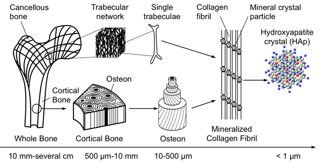
Fig. 1. Hierarchical structure of bone
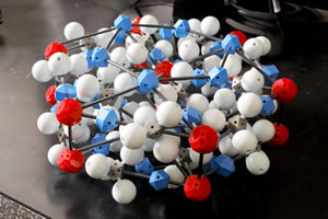
Photo 1. Hydroxyapatite crystal molecule model
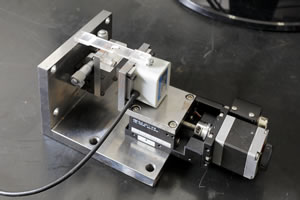
Photo 2. Small mechanical testing device created in the laboratory